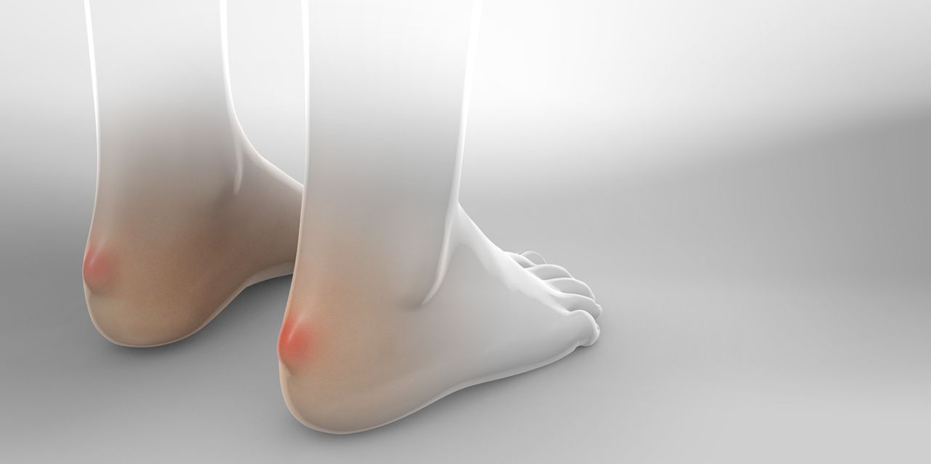
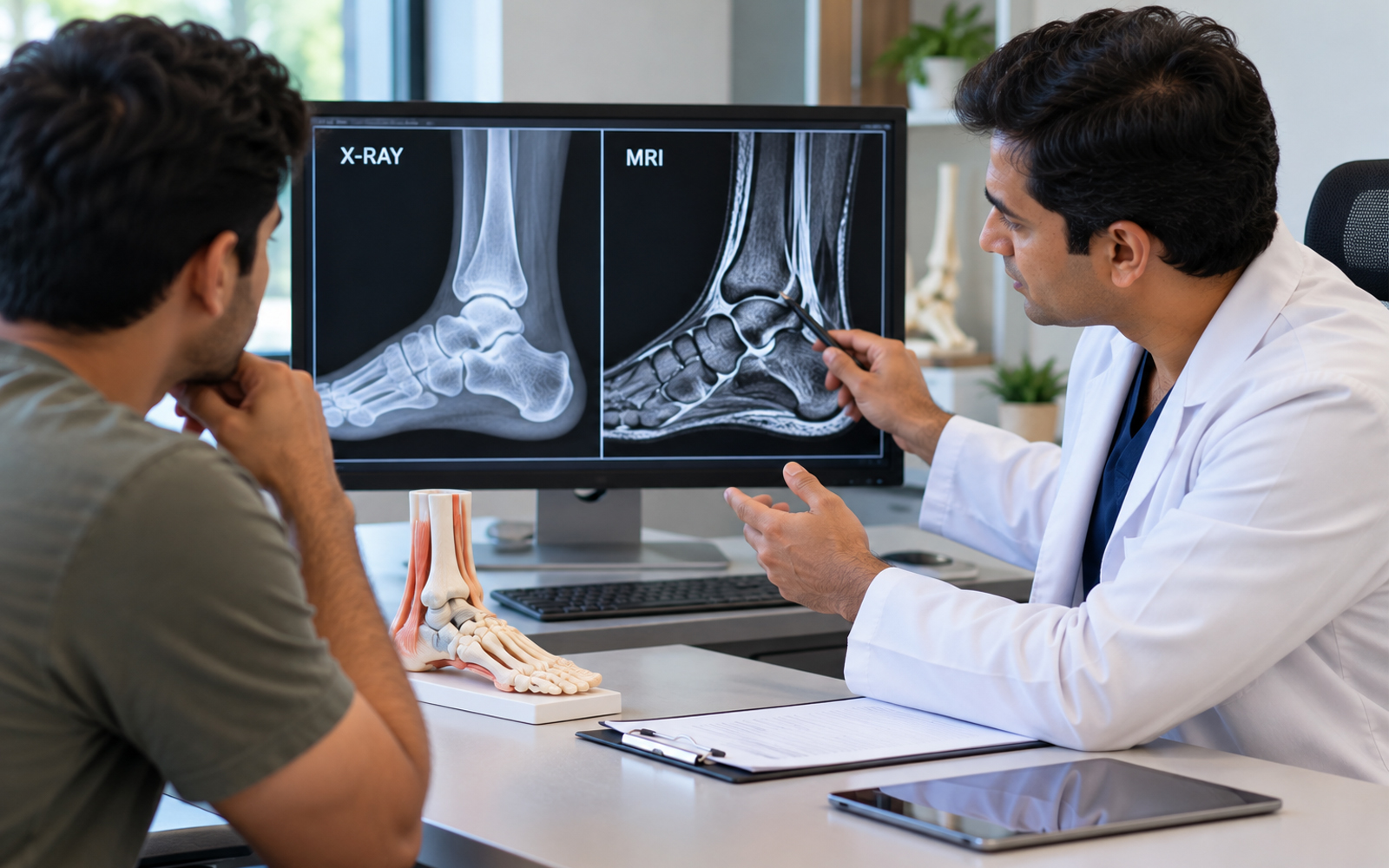
If you're dealing with foot or ankle pain, one of the first questions your doctor will answer isn't "what's wrong" it's "what scan do we need to find out." X-ray and MRI are both used for foot and ankle problems, but they show completely different things, and ordering the wrong one first can delay a proper diagnosis by weeks.
This guide breaks down exactly what each test does, when each one is actually necessary, and which imaging is typically used for common foot and ankle conditions from a simple ankle sprain to more complex problems like Charcot foot or adult acquired flatfoot.
What Does a Foot & Ankle X-Ray Show?
An X-ray uses radiation to create a 2D image of bone. It's fast, inexpensive, and it's almost always the first test ordered for any foot or ankle complaint.
X-rays are good at showing:
- Fractures and breaks, including stress fractures once they've matured.
- Bone alignment and deformity like the angle of a bunion (hallux valgus) or the position of a hammer toe.
- Joint space narrowing, which points to arthritis.
- Bone spurs, such as a heel spur associated with plantar fasciitis.
- Overall foot structure, arch height, forefoot alignment, and signs of flat foot or progressive collapsing foot deformity.
- Hardware position after a previous surgery, including fusion, ankle replacement, or osteotomy.
What an X-ray cannot show: soft tissue. Ligaments, tendons, cartilage, nerves, and early bone marrow changes are essentially invisible on a plain X-ray. This is the single most common reason X-ray results come back "normal" even though a patient is clearly in pain the injury is in the soft tissue, not the bone.
What Does a Foot & Ankle MRI Show?
An MRI uses magnetic fields, not radiation, to produce detailed cross-sectional images of both bone and soft tissue. It takes longer and costs more, so it's rarely the first test it's ordered when an X-ray doesn't explain the symptoms, or when the suspected problem is soft-tissue in nature from the start.
MRI is the test of choice for:
- Ligament tears, ankle sprains that aren't healing, or suspected ankle instability.
- Tendon problems, including Achilles tendon injury and posterior tibial tendon damage, a major cause of adult acquired flatfoot.
- Nerve compression, including Morton's neuroma, which is a classic MRI diagnosis rather than an X-ray diagnosis.
- Cartilage damage inside the ankle joint.
- Bone stress injuries that are too early to show up on X-ray, including early stress fractures.
- Occult or hidden fractures, especially in the midfoot. This is critical for suspected Lisfranc injury, which X-rays frequently miss in its early or subtle form.
- Infection and bone involvement, used to assess diabetic foot ulcer complications and Charcot foot activity.
- Soft tissue masses, chronic tendon degeneration, or planning before ligament reconstruction or tendon repair surgery.
When Do You Actually Need an X-Ray First?
- Any acute injury with swelling, deformity, or inability to bear weight.
- Suspected fracture after a fall, twist, or direct trauma.
- Chronic bunion or hammer toe evaluation, to measure deformity severity before considering bunion surgery or hammer toe correction.
- Persistent heel pain to rule out a heel spur before starting plantar fasciitis treatment.
- Follow-up after any foot reconstruction surgery, to confirm bone healing and hardware position.
When Does Your Doctor Move to MRI?
Your foot and ankle specialist will typically order an MRI when:
- The X-ray is normal but pain, swelling, or instability persists beyond 2-3 weeks.
- A ligament or tendon injury is suspected, for example, recurrent ankle sprains raising concern for ankle instability that may need ligament reconstruction.
- Nerve-type symptoms are present, including burning, tingling, or a "pebble in my shoe" sensation, the hallmark of Morton's neuroma or metatarsalgia.
- There's concern about a Lisfranc injury, since these midfoot ligament injuries are notoriously easy to miss on X-ray alone.
- A diabetic patient has a non-healing foot ulcer or suspected Charcot foot, where MRI helps distinguish infection from bone remodeling, which is critical for deciding on limb salvage surgery versus more conservative foot infection treatment.
- Surgery is being planned. MRI is often used pre-operatively for flatfoot reconstruction surgery, ankle arthroscopy, or cartilage repair in the ankle.
Why the Right Scan Matters Before Treatment
Getting the correct imaging early isn't just about diagnosis speed — it directly shapes treatment decisions. A patient with a suspected ankle ligament injury who only gets an X-ray may be told "nothing is broken" and sent home, when an MRI would have shown a tear needing ankle ligament reconstruction. Similarly, subtle Lisfranc injuries missed on X-ray can progress to painful midfoot arthritis if not caught early with MRI.
This is why, at Dr. Harsh Vyas's clinic, imaging is matched to the suspected condition rather than defaulted to "one test for everything" whether the path ahead involves conservative care, an injection (such as a PRP injection for foot pain or hyaluronic acid injection for the ankle), or surgical correction such as:
- Bunion Surgery / Hallux Valgus Correction
- Hammer Toe Correction
- Flatfoot Reconstruction Surgery / Calcaneal Osteotomy
- Ankle Ligament Reconstruction / Ankle Ligament Repair
- Ankle Arthroscopy / Arthroscopic Ankle Surgery
- Ankle Fusion Surgery / Ankle Replacement Surgery
- Achilles Tendon Repair
- Limb Salvage Surgery for Diabetic Foot